产品

本公司产品仅供体外研究使用,不用于临床诊断

anti- ELKS antibody

FNab10262 货号
别称:
ELKS, KIAA1081, RAB6IP2, ERC1
宿主:
Rabbit
适应种属:
Human, Mouse, Rat
亚型:
IgG
验证应用:
ELISA, WB
产品信息
产品名称
anti- ELKS antibody
货号
FNab10262
规格
100µg
状态
liquid
纯化方法
Immunogen affinity purified
纯度
≥95% as determined by SDS-PAGE
宿主
Rabbit
克隆性
polyclonal
亚型
IgG
存储
PBS with 0.02% sodium azide and 50% glycerol pH 7.3,-20℃ for 24 months(Avoid repeated freeze / thaw cycles.)
背景介绍
Regulatory subunit of the IKK complex. Probably recruits IkappaBalpha/NFKBIA to the complex. May be involved in the organization of the cytomatrix at the nerve terminals active zone(CAZ) which regulates neurotransmitter release. May be involved in vesicle trafficking at the CAZ. May be involved in Rab-6 regulated endosomes to Golgi transport.
免疫原信息
免疫原
ELKS/RAB6-interacting/CAST family member 1
别称
ELKS, KIAA1081, RAB6IP2, ERC1
分子量
130 kDa,110 kDa, 82 kDa
应用
验证应用
ELISA, WB
建议稀释比例
WB: 1:500 - 1:2000
Uniprot 信息
UniProt ID
Q8IUD2
图像
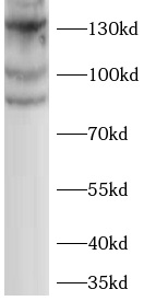
NIH/3T3 cells were subjected to SDS PAGE followed by western blot with FNab10262(ELKS antibody) at dilution of 1:1000

Experimental Procedure of Primary Antibody

Protocol for Immunoblotting and Immunoprecipitation:

Preparation of Cell Lysate:

Add RIPA buffer to cells(100μl to a 35mm dish, 200μl to a 60 mm dish, 500 uL to a 100 mm dish) while the culture dish is placed on ice. Scrape the cells and gently rock the suspension on either a rocker or an orbital shaker in the cold room for 15 minutes to lyse cells. Sonicate in ice water with bath sonicator, until the sample is no longer viscous. Centrifuge the cells at 12000 g at 4°C for 5 minutes to remove pellet. Move the supernatant to a fresh tube. Final concentration of cell lysate will be 2-3 ug/uL. Add 5 x SDS stop buffer to the lysate to a 1xSDS final concentration .

Immunoblotting:

  1. After boiling for 10 minutes, 50-80 uL sample will be loaded to SDS PAGE. For most proteins, 100-200 ug total lysate proteins per lane loading amount is suggested, because 80% of protein species in cells are at very low concentration.
  2. Soak the gel in western blot transfer buffer. Cut a piece of membrane to the size of the gel. PVDF membrane is recommended for most proteins. Dip the membrane into methanol for 1-2 minutes, soak the membrane in transfer buffer for 10 minutes, and place it on a thick stack of buffer-soaked filter paper. Then cover it with another stack of buffer soaked filter papers. Cover up the transfer apparatus. Gel should be on the negative side of the membrane.
  3. Run for 90 minutes at current of 1 mA per cm2 .
  4. Incubate the membrane in blocking buffer for 1 hour at room temperature or overnight at 4°C. Wash membrane 3x5 min with washing buffer.
  5. Dilute primary antibody with blocking buffer. Incubate the membrane for 1 hour at room temperature or overnight at 4°C. Wash membrane 3x5 min with washing buffer.
  6. Dilute secondary antibody with blocking buffer. Incubate the membrane for 1 hour at room temperature. Wash membrane 3x5 min with washing buffer.
  7. Wash membrane 3x10 min with washing buffer.
  8. Develop color with ECL.

Immunoprecipitation:

  1. 1. Prepare cell lysates or other protein samples. Cellular proteins or other protein samples should be labeled either metabolically or by iodination or biotination. For cell lysate, a concentration of 1-2 ug/uL protein is optimum.
  2. To 500 ug - 1 mg cell lysate, add 50 uL of 50% slurry of protein A-sepharose beads and incubate the mix at 4°C for 15 minutes. Centrifuge at 10000 g for 5 minutes at 4 °C. Collect the supernatant.
  3. To the supernatant add 5 ug of rabbit polyclonal antibodies against the specific antigen. Gently rock the reaction mixture for 1 hour at 4 °C.
  4. Add 50 uL of 50% slurry of protein A-Sepharose beads to the reaction mixture and gently rock the sample at 4°C for 30 minutes.
  5. Spin the sample at 10000 g at 4°C for 3 minutes to pellet the beads.
  6. Wash the beads twice with washing buffer A (10 mM Tris-HCl, pH 8, 500 mM NaCl, 0.5% NP-40 and 0.05% SDS), once with washing buffer B (10 mM Tris-HCl, pH 8, 150 mM NaCl, 0.5% NP-40, 0.05% SDS and 0.5% Dideoxycholate), once with washing buffer C (10 mM Tris-HCl, pH 8, 0.05% SDS).
  7. Add 100 uL of 1.25 x SDS loading buffer and boil the sample for 10 minutes before loading to SDS PAGE.
  8. Detect the immunoprecipiated sample on SDS-PAGE either by direct radioautography or by further blotting biotin, depending on the initial labeling method.

Protocol for Immunofluorescence with Tissue Culture Cells

  1. Culture the cells on small round cover glass.
  2. Fix cells with formalin-PBS (10%) for 10 minutes at 4°C.
  3. Transfer the glass into pre-chilled acetone (-20°C) for exact 5 minutes.
  4. Transfer the cover glass into PBS, the cells on cover glass can be stored in PBS-NaN3 for several months at 4°C. 5. Block the cells with BSA-PBS-NaN3.
  5. Add 40 uL of specific primary antibodies at concentration of 1 ug/mL on cover glass and incubate for 1 hour at room temperature in a moist container in dark.
  6. Wash the cover glass with 6 cups of PBS-N aN3.
  7. Add 40 uL of fluorescence-labeled secondary antibody on each cover glass and incubate for 1 hour at room temperature in a moist container in dark.
  8. Mount the coverslip in 15% glycerol or a small volume of hydromount on a slide.
  9. Seal the coverslip with nail polish on the edge.

Protocol for Immunofluorescence with frozen tissue section

  1. The frozen sections were removed and balanced at room temperature for 10-20min. The sections could not be completely dried.
  2. Fix with 4% paraformaldehyde at room temperature for 15min, then rinse 3 x 5 min with TBS.
  3. Place section in vessel filled with 3% H2O2 solution at room temperature for 10min. The solution must be prepared freshly .The preparation method of 3% H2O2 solution is to add 1ml 30% H2O2 to 9ml methanol, mix completely.
  4. Rinse 3 x 5 min with TBS.
  5. Block in 5% BSA solution for 30 minutes at 37℃.
  6. Drain slides for a few seconds (do NOT rinse) and wipe round sections.
  7. Add 40 uL of specific primary antibodies at concentration of 1 ug/mL on cover glass and incubate for 2 hour at 37℃ or overnignt at 4℃ in a moist container in dark.
  8. Rinse 1 x 5 min with TBST. Then rinse 3 x 5 min with TBS.
  9. Add 40 uL of fluorescence-labeled secondary antibody on each cover glass and incubate for 1 hour at room temperature in a moist container in dark.
  10. Rinse 1 x 5 min with TBST. Then rinse 3 x 5 min with TBS.
  11. Add DAPI or PI working solution to cover glass and incubate for 10min at room temperature in dark.
  12. Rinse 3 x 3 min with TBS.
  13. Mount the coverslip in 15% glycerol or a small volume of hydromount on a slide.
  14. Seal the coverslip with nail polish on the edge.

Protocol for Immunohistochemistry on Paraffin-Embedded Tissue Sections

  1. Deparaffinize slides in 2 changes of xylene, one for 40 minutes, the other for 20 minutes.
  2. Transfer slides to 100% alcohol, 95% alcohol, 80% alcohol, 60% alcohol and ddH2O for 5 minutes each.
  3. Place slides in vessel filled with antigen retrieval buffer and microwave on middle for about five minutes (700 W oven), allow retrieval solution to cool at room temperature.
  4. Rinse 2 x 5 min with TBS.
  5. Block endogenous peroxidase activity by incubating sections in 3% H2O2 solution for 30 minutes.
  6. Rinse 2 x 5 min with TBS.
  7. Block in 5-10% goat serum in TBS for 30 minutes at room temperature.
  8. Drain slides for a few seconds (do NOT rinse) and wipe round sections.
  9. Apply primary antibody made up in TBS. Incubate 1 hour at room temperature or 4°C overnight.
  10. Rinse 2 x 5 min with TBST.
  11. Apply secondary biotinylated antibody made up in TBS for 1 hour at room temperature.
  12. Rinse 2 x 5 min with TBS.
  13. Apply streptavidin peroxidase for 15 minutes or 30 minutes at room temperature.
  14. Rinse 2 x 5 min with TBS.
  15. Develop with chromogen (DAB) at room temperature, watching under microscope.
  16. Rinse in running tap water for 5 minutes.
  17. Counterstain in mayor’s hematoxylin bath for 30-60 seconds.
  18. Wash in water bath 7-8 times, then tap water for 3 minutes.
  19. Dehydrate through 60%, 80%, 95% and 100% alcohol for 5 minutes each. 20. Transfer to xylene for 5 minutes. Air for 30 minutes.
  20. Mounting.

Buffers

10 x PBS 2000 mL Transfer buffer 1000 mL BSA-PBS-NaN3 1000 mL

H2O 1000 mL Tris base 48 mM BSA 5 g

KCl 4 g Glycine 39 mM 1 x PBS-Azide 1000 mL

KH2PO4 4 g SDS 0.037%

Na2HPO4· H2O 22.9 g Methanol 20%

NaCl 160.0 g

ddH2O up to 2 liter,

Filter and Autoclave

Washing buffer TBST pH 7.4 RIPA buffer 1000 mL Blocking buffer

10 mM Tris-HCl Sodium Chloride 150 mM Non-fat Milk 5 g

100 mM NaCl NP-40 or Triton X-100 1.0% Washing buffer TBST 100mL

0.2% Tween-20 Sodium Deoxycholate 0.5%

SDS 0.1% PBS-NaN3 1000 mL

TBS-Triton X-100 1000 mL Tris base 50 Mm 1 x PBS 1000 mL

TBS 1000 mL EGTA 1 mM Na-Azide 2 g

Triton X-100 250 uL EDTA 5 mM

Sodium Fluoride 5 -10 mM

Sodium Orthovanadate 1 mM

Antigen retrieval buffer 500 mL TBS 1000 mL

0.1 M Citric Acid 9 mL Tris base 3 g

0.1 M Sodium Citrate 41 mL NaCl 8 g

ddH2O 450 mL ddH2O 1000 mL

pH 6.0 Adjust pH to 7.6 with HCl

Immunohistochemistry Q&A

Technically, IHC and ICC are relatively simple and straightforward experimental methods. However, there are many variables which must be identified and optimized for each individual IHC/ICC study. Optimization of IHC/ICC may also require troubleshooting a variety of factors. The tables below highlight common IHC/ICC issues and provide appropriate experimental actions. For further assistance, please contact our technical service department.

Problem: Lack of Staining

Possible SourceSuggestion
Lack of antigen.Check protein expression by in situ hybridization (in some rare cases translation may be blocked even though mRNA is detected).
Antibodies do not work due to improper storage.Follow storage instructions on the datasheet. In general, aliquot antibodies into smaller volumes sufficient to make a working solution for a single experiment. Store aliquots in a manual defrost freezer (-20 to -70 °C) and avoid repeated freeze-thaw cycles.
Inactive primary or secondary antibodies.Test reporter system independently to assess reagent viability.
Inadequate tissue fixation.Try increasing the fixation time or try a different fixative.
Tissue overfixation.Reduce the duration of the immersion or post-fixation steps. If immersion fixation cannot be avoided (for example, collection of postmortem tissues or biopsies in pathology lab), antigens may be unmasked by treatment with antigen retrieval reagents.
Incompatible secondary and primary antibodies.Use a secondary antibody that will interact with the primary antibody. For example, if the primary antibody was raised in rabbits, use an anti-rabbit secondary antibody.
Antigen was destroyed before incubation with the primary antibody.If quenching of endogenous peroxidase was done prior to the addition of primary antibodies, block peroxidase after incubation with the primary antibody.
Epitope altered during fixation or embedding procedure.Try restoring immunoreactivity through various antigen retrieval techniques.
Embed tissue at 58 °C or below.
Antigen retrieval was ineffective.Increase the time of treatment or change the treatment solution.
Reagents omitted or used in wrong order.Repeat staining and confirm that correct reagents are used and are added in the correct order.

Problem: High Background

Possible SourceSuggestion
High concentration of primary and or secondary antibodies.Titer antibody to determine optimal concentration needed to promote a specific reaction of the primary and the secondary antibodies.
Hydrophobic interactions of the antibody and proteins in the tissue.Lower the ionic strength of the antibody diluent (particularly monoclonal antibodies respond well to reducing the salt concentration).
Non-speci?c binding of primary and/or secondary reagents to tissues.Use blocking step just prior to primary antibody incubation (we use 1% bovine serum albumin with 10% normal donkey serum). Non-fat dry milk is another option.
Non-specific binding of secondary antibody.Use an antibody that has cross-reactive IgG species removed (absorbed against sample species).
Tissue dried out.Avoid letting the tissue dry during the staining procedure.
Reagents sticking to old or poorly prepared slides.Start over with freshly prepared or purchased slides.
Background due to ionic interactions.Increase the ionic strength of the diluent buffer.

Problem: Cell/Tissue Morphology is Destroyed

Possible SourceSuggestion
Antigen retrieval methods are too harsh.Empirically determine the conditions that preserve tissue morphology while restoring the immunoreactivity of the antigen.
Tissue sections falling off slide. (more common with frozen sections)Increase fixation time. Empirically determine an additional or alternative fixative.
Use freshly prepared, adequately charged slides.
Tissue section appears torn or folded. Air bubbles under section.Re-cut sections using a sharp blade, or ignore damaged areas when analyzing the results.
Poor resolution of tissue morphologyCut thinner tissue sections. Ice crystals may have destroyed morphology of frozen sections.
Underfixation has physically damaged the tissue or cells during the staining processIncrease fixation time and/or add a post-fixation step. Increase the fixative/tissue ratio.
Cut smaller pieces of tissue for more thorough immersion fixation.
Autolysis of tissue leading to staining of necrotic debris.Increase the fixation time, ratio. Consider using cross-linking fixative.

Problem: Staining is Inappropriate

Possible SourceSuggestion
Fixation method is inappropriate for the antigen.Try a different fixative or increase the fixation time.
Antigen retrieval may be inappropriate for this antigen or tissue.Try different antigen retrieval conditions.
Electrostatic charge of the antigen has been altered.Try adjusting the pH or cation concentration of the antibody diluent.
Delay in fixation caused diffusion of the antigen.Fix tissue promptly. Try a cross-linking fixative rather than organic (alcohol) fixative.

Western Blot Q&A

For further assistance, please contact our technical service department.

Problem: No Bands Observed

Possible SourceSuggestion
Insufficient antibodyAntibody may have low affinity to protein of interest. Increase antibody concentration (2-4 fold higher than recommended starting concentration).
Antibody may have lost activity. Perform a Dot Blot.

Insufficient protein

Increase the amount of total protein loaded on gel.
Confirm the presence of protein by another method.
Use a positive control (recombinant protein, cell line or treat cells to express analyte of interest).
Perform a Dot Blot.

Poor transfer

Wet PVDF/Immobilon-P membrane in methanol or nitrocellulose membrane in transfer buffer.
Ensure that there is good contact between PVDF membrane and gel.

Incomplete transfer

Optimize transfer time. High MW protein may require more time for transfer.
To ensure transfer is complete, stain the membrane with Ponceau S, Amido Black or India Ink.
Use prestained MW marker.
Over transferReduce voltage or time of transfer for low molecular weight proteins (< 10 kDa).
Isoelectric point is >9Use alternative buffer system with higher pH such as CAPS (pH 10.5).
Incorrect secondary antibody usedConfirm host species and Ig type of primary antibody.
Old antibodyIf antibody is expired or past manufacturer warranty, purchase fresh antibody.
Incorrect storage of antibodiesFollow manufacturer's recommended storage and avoid freeze/thaw cycles.
Sodium Azide contaminationMake sure buffers do not contain Sodium Azide as this can quench HRP signal.
Insufficient incubation time with primary antibodyExtend incubation time to overnight at 4°C.

Problem: Faint Bands (Weak Signal)

Possible SourceSuggestion

Low protein-antibody binding

Reduce the number of washes to minimum.
Reduce NaCl concentration in Blotting Buffer used for wash steps (recommended range 0.15M - 0.5M).
Reduce NaCl concentration in Antibody Solution (recommended range 0.15M - 0.5M).
Insufficient antibodyAntibody may have low affinity to protein of interest. Increase antibody concentration (2-4 fold higher than recommended starting concentration).
Insufficient proteinIncrease the amount of total protein loaded on gel.
Inactive conjugateMix enzyme and substrate in a tube. If color does not develop or, it is weak. Make fresh or purchase new reagents. Switch to ECL.
Weak/Old ECLPurchase new ECL reagents.
Non-fat dry milk may mask some antigenDecrease milk percentage in Block and Antibody Solutions or substitute with 3% BSA.

Problem: Extra Bands

Possible SourceSuggestion

Non-specific binding of primary antibody

Reduce primary antibody concentration.
Reduce the amount of total protein loaded on gel.
Use monospecific or antigen affinity purified antibodies.

Non-specific binding of secondary antibody

Run a control with the secondary antibody alone (omit primary antibody). If bands develop choose an alternative Secondary Antibody.
Use monospecific or antigen affinity purified antibodies.

Non-specific binding of primary or secondary antibodies

Add 0.1 - 0.5% Tween20 to primary or secondary Antibody Solution.
Use 2% non-fat dry milk in Blotting Buffer as a starting point to dilute primary and secondary antibodies. Adjust antibody concentration up or down as needed.
Increase number of washes.
Increase NaCl concentration in Blotting Buffer used for antibody dilution and wash steps (recommended range 0.15M - 0.5M).
Increase Tween20 concentration in Blotting Buffer used for wash steps (0.1%-0.5%).

Aggregation of analyte

Increase amount of DTT to ensure complete reducing of disulfide bonds (20 -100mM DTT). Heat in boiling water bath 5-10 minutes before loading onto gel.
Perform a brief centrifugation.

Degradation of Analyte

Minimize freeze/thaw cycles of sample.
Add protease inhibitors to sample before storage.
Make fresh samples.
Contamination of reagentsCheck buffers for particulate or bacterial contamination. Make fresh reagents.

Problem: High Background

Possible SourceSuggestion

Non-specific binding of primary antibody

Use monospecific or antigen affinity-purified antibodies.
Block in 5% milk. Adjust the milk (2-5%) or NaCl (0.15-0.5M) concentrations of primary Antibody Solution.
Decrease antibody concentration.
Non-specific binding of secondary antibodyRun a control with the secondary antibody alone (omit primary antibody). If bands develop choose an alternative Secondary Antibody.

Insufficient blocking

Start with 5% dry milk with 0.1%- 0.5% Tween 20, 0.15 -0.5M NaCl in 25mM Tris (pH 7.4). Incubation time may be extended. Adjust milk concentration up or down as needed.
Overnight blocking at 4°C may decrease blocking efficiency since detergents might not be effective at lower temperatures.
Non-fat dry milk may contain target antigenSubstitute with 3% BSA.
Non-fat dry milk contains endogenous biotin and is incompatible with avidin/streptavidinSubstitute with 3% BSA.
Some IgY antibodies may recognize milk proteinSubstitute with 3% BSA.

Insufficient wash

Increase number of washes.
Increase Tween 20 concentration in Wash Buffer (0.1%-0.5%).
Non-Specific Binding of Primary AntibodyIncrease NaCl concentration in primary Antibody Solution and Blotting Buffer used for dilution of primary antibody and wash steps (recommended range 0.15M - 0.5M).

Film overexposed

Reduce exposure time.
If target signal is too strong wait 5-10 minutes and re-expose to film.

Problem: Diffuse Bands

Possible SourceSuggestion
Excessive protein on gelReduce amount of protein loaded

Problem: White Bands (ECL method)

Possible SourceSuggestion
Excessive signal generatedReduce antibody or protein concentration. Excessive antibody or protein can cause extremely high levels of localized signal (usually at a single band). This results in rapid, complete consumption of substrate at this point. Since there is no light production after the completion of this reaction, white bands are the result when exposed to film.

Problem: Patchy uneven spots all over the blot

Possible SourceSuggestion
Contamination of reagentsCheck buffers for particulate or bacterial contaminate. Make fresh reagents.
Not enough solution during incubation or washingMake sure membrane is fully immersed during washes and antibody incubations.
Air bubble trapped in membraneGently remove any air bubbles. Especially during transfer.
Uneven agitation during incubationsEnsure uniform agitation by placing on a rocker/shaker.
Contaminated equipmentMake sure that the electrophoresis unit is properly washed. Protein or pieces of gel remaining on the unit may stick to the membrane. Wash membrane thoroughly.
HRP aggregationFilter conjugate to remove HRP aggregates.
Long exposureReduce exposure time.